Advancing Expansion Microscopy Techniques for Plant Research
Advancing Expansion Microscopy Techniques for Plant Research
In an exciting advancement in plant biology, researchers at Washington University in St. Louis have introduced a groundbreaking technique known as ExPOSE, short for Expansion Microscopy in Plant Protoplast Systems. This innovative method adapts the already established expansion microscopy for studies involving plant cells, thereby transforming the conventional ways of observing these vital biological units. Kevin Cox, an assistant professor of biology in Arts & Sciences at the university, and his dedicated team have broken new ground in plant imaging, which is pivotal for understanding cellular mechanisms and communication within plant tissues.
The traditional approaches to imaging biological samples, especially in plant research, come with inherent limitations. Conventional optical microscopes offer various degrees of resolution and usability. Low-end models tend to sacrifice detail for user-friendliness, producing images that lack the necessary clarity to understand cellular structures thoroughly. On the other hand, high-end microscopes provide exceptional resolution but are often complicated and expensive, presenting a barrier for many researchers in the field. The introduction of ExPOSE presents a solution that allows researchers to overcome these trade-offs by offering a method that enables them to visualize plant cells with unprecedented clarity and detail, all while maintaining accessibility.
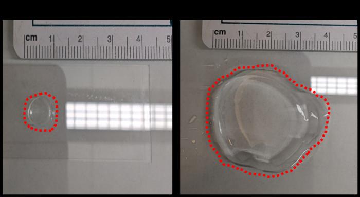
The central principle of ExPOSE is the use of a hydrogel—a water-absorbing polymer that expands without distorting its shape—to physically enlarge biological tissues. This innovative technique stands in stark contrast to traditional microscopy that relies on optical zooming, which often leads to blurriness and distortion. By embedding plant cells in this hydrogel, researchers can achieve higher resolution images that showcase cellular components more effectively. As the hydrogel swells, the embedded cellular structures also expand, akin to a sponge absorbing water. This unique approach not only enhances visualization but does so at a fraction of the cost associated with high-end imaging technologies.
While the application of expansion microscopy has flourished within animal research, applying this pioneering technique to plant studies has not been straightforward. The primary hurdle arises from the rigid cell walls typical of plant cells, composed of cellulose. These cell walls inhibit the uniform expansion needed for effective imaging. Cox and his team addressed this challenge by utilizing plant protoplasts—cells with their outer walls removed—thereby achieving the uniformity necessary for successful expansion microscopy in plant systems. The successful adaptation and implementation of ExPOSE marks a significant milestone that could pave the way for a deeper understanding of plant cell structures.
With ExPOSE, researchers can now visualize intricate details within plant cells, enabling enhanced studies of proteins, RNA, and various biomolecules. Understanding the precise localization of these cellular components is critical, especially for researchers like Cox, whose work revolves around cellular communication and responses. By examining how these biomolecules are positioned within plant structures, scientists gain valuable insights into their functions and implications in broader plant physiology.
Moreover, the versatility of ExPOSE can be augmented by coupling it with other imaging techniques. When combined with hybridization chain reaction (HCR) and immunofluorescence, researchers have observed even greater resolution and detail in their images, allowing for multidimensional analyses of plant biology. This amalgamation of methodologies lays the foundation for a comprehensive toolkit that can significantly advance the accessibility of complex biological studies.
Although current investigations employing ExPOSE focus on single cell imaging, Cox harbors aspirations for expanding this technique to encompass larger biological scales. He envisions utilizing ExPOSE to analyze entire plant organs, leaves, and even whole plants. This comprehensive approach could revolutionize our understanding of cellular communication not only within individual cells but also among the broader plant structure. By decoding these cellular interactions, researchers can uncover how plants coordinate responses to environmental challenges and intercellular signaling.
A notable advantage of Cox’s research is the use of duckweed as a model organism. This small, rapidly growing aquatic plant can serve as a powerful tool for exploring cellular communication and gene expression. Duckweed’s diminutive size enables researchers to scrutinize cellular behavior in real time, presenting an unparalleled opportunity to gain insights into how individual cells react to stressors such as disease or environmental fluctuations. By mastering the intricacies of cellular responses within duckweed, scientists aspire to translate their findings to crops, ultimately contributing to advances in agriculture.
Enhancing our understanding of cellular interactions in plant species can lead to revolutionary advancements in agricultural practices. By clarifying how plant cells communicate and defend themselves against stressors, researchers can work towards developing resilient crops that yield more produce while also withstanding environmental fluctuations. This knowledge is increasingly crucial in a world where food security and sustainability become pressing challenges.
As ExPOSE continues to gain traction in the field of plant research, the implications of this technique extend far beyond academic inquiry. The potential to develop crops that respond more effectively to environmental changes could enhance food security worldwide. By harnessing these insights, agricultural science could make significant strides in crafting crops capable of thriving in a fluctuating climate, thereby ensuring that global food needs are met efficiently.
The scientific community eagerly anticipates further developments stemming from this innovative technique. The intersection of molecular biology and imaging technologies offers a fertile ground for discovering previously uncharted realms of plant biology. As researchers begin to realize the transformative potential of ExPOSE, collaboration across disciplines will likely drive forward not only the understanding of plant biology but also the advancement of sustainable agricultural solutions.
In summary, the introduction of ExPOSE presents a remarkable leap in plant research techniques. By utilizing expansion microscopy in protoplast systems, scientists unlock impressive opportunities to visualize and understand intricate cellular structures and interactions. With the potential for far-reaching agricultural applications and the promise of enhanced research capabilities, ExPOSE is poised to become an indispensable tool in the quest to unlock the secrets held within plant biology.
Subject of Research: Expansion Microscopy in Plant Protoplast Systems
Article Title: ExPOSE: a comprehensive toolkit to perform expansion microscopy in plant protoplast systems
News Publication Date: March 6, 2025
Web References: Washington University in St. Louis
References: The Plant Journal
Image Credits: Cox Laboratory, Washington University in St. Louis
Keywords
Life sciences, Plant sciences, Expansion microscopy, Hydrogel, Plant biotechnology, Cellular communication, Plant protoplasts, Plant genetics, Agriculture, Food crops.
Emerging Innovators Shine at UTA Science Fair
Next PostNew Initiative Aims to Enhance Cancer Gene Testing in Primary Care Settings
Related Posts

Random-Event Clocks Offer New Window into the Universe’s Quantum Nature
Scientists reinvigorate pinhole camera technology for advanced next-generation infrared imaging
Portable Light-Based Brain Monitor Demonstrates Potential for Advancing Dementia Diagnosis
BeAble Capital Invests in UJI Spin-Off Molecular Sustainable Solutions to Advance Disinfection and Sterilization Technologies
Scientists Pioneer Innovative Method for Precise Experimental Measurement of the Unruh Effect